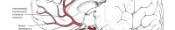


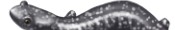


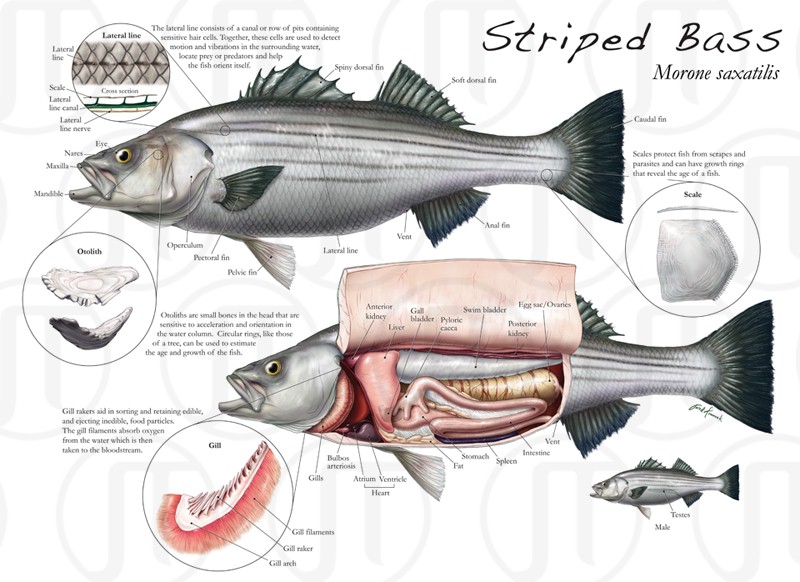
External and Internal Anatomy of Morone saxatilis
The Morone saxatilis, or Striped Bass, is an essential element in the Chesapeake bay ecosystem. Grade students learn about the importance of this fish through a course at the National Aquarium in Baltimore. This illustration was created to help guide the students through the fish’s external and internal anatomy. This illustration received the 2009 AMI Award of Excellence.
Text: The lateral line consists of a canal or row of pits containing sensitive hair cells. Together, these cells are used to detect motion and vibrations in the surrounding water, locate prey or predators, and help the fish orient itself. Otoliths are small bones in the head that are sensitive to acceleration and orientation in the water column. Circular rings, like those of a tree, can be used to estimate the age and growth of the fish. Gill rakers aid in sorting and retaining edible and ejecting inedible food particles. The gill filaments absorb oxygen from the water which is then taken into the bloodstream. Scales protect fish from scrapes and parasites and can have growth rings that reveal the age of a fish.
Anatomy in this biological illustration: Lateral line, Scale, Lateral line canal, Lateral line nerve, Eye, Nares, Maxilla, Mandible, Otolith, Operculum, Pectoral fin, Pelvic fin, Vent, Anal fin, Scale, Caudal fin, Soft dorsal fin, Spiny dorsal fin, Gill, Gill filaments, Gill raker, Gill arch, Bulbos arteriosis, Heart, Atrium, Ventricle, Fat, Stomach, Spleen, Intestine, Testes