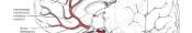

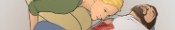
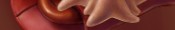
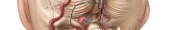
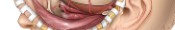
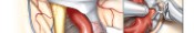


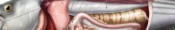

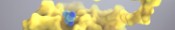


Nissen Fundoplication with Cruroplasty
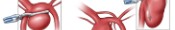
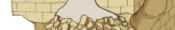
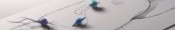
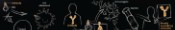
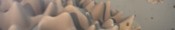
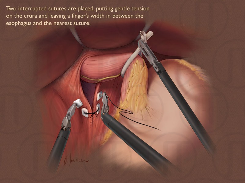
The stomach and esophagus have been removed from the chest cavity and the esophageal hiatus is being sutured shut using the DaVinci laparoscopic robot (Figure 01). Figures 02 and 03 were sketches produced to show the steps in the entire procedure. This illustration received the 2009 AMI Award of Merit.
Anatomy in this medical illustration: Diaphragm, Esophagus, Stomach, Liver, Posterior vagul trunk, Anterior vagul trunk, Vagus nerve, Esophageal hiatus, Crura, Esophagogastric fat pad, Gall baldder, Left gastric artery, Right gastric artery, left gastroomental artery, Common hepatic artery, Proper hepatic artery


