


The Creation of a Medical Illustration
Watch my process of creating a complex medical illustration in this video created for Fortnight Journal.
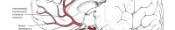
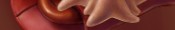
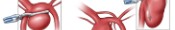
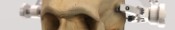
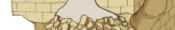
This is the 4th illustration in a set of 10 that describes the clipping of a posterior inferior cerebellar artery aneurysm. In this illustration the craniotomy and C1 laminectomy have been performed and a rongeur is removing the remaining portion of the skull base to the foramen magnum.
This video is one of six posts in a series by me about the profession of medical illustration from the vantage point of the Millennial generation. Visit www.fortnightjournal.com to see more.
To see my other articles browse the links below:
http://fortnightjournal.com/jared-travnicek/265-the-drawn-body.html
http://fortnightjournal.com/jared-travnicek/279-a-scientist-renders.html
http://fortnightjournal.com/jared-travnicek/293-the-future.html
http://fortnightjournal.com/jared-travnicek/309-street-anatomy.html
http://fortnightjournal.com/jared-travnicek/323-on-copyright.html
http://fortnightjournal.com/jared-travnicek/334-my-process.html
Anatomy in this medical illustration: Foramen magnum, Vertebral artery, Verterbral artery venous plexus, Cervical bone, C1, Skull, Transverse sinus, Sigmoid sinus, Mastoid process, Occipital bone, Fat, Muscle, Craniotomy, Laminectomy, Dura, Posterior inferior cerebellar artery aneurysm, Skull base